
You may have been diagnosed with skin cancer and were told the best treatment option is Mohs Micrographic surgery. If you want to learn more about Mohs surgery you have come to the right place.
What is Mohs Surgery?
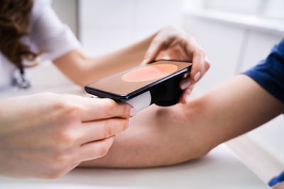
Let’s start from the beginning … Skin cancer may initially appear as a bump or irregular patch on the surface of the skin. As the cancer grows, the size or shape of the visible skin mass may change. Some skin cancers can be deceptively large, sending out “roots” and extensions that are far more extensive under the skin than they appear to be from the surface. If left untreated, the skin cancer may spread into nearby muscle tissue, blood vessels, nerves, cartilage, and depending on the type of skin cancer, lymph nodes and other organs.
Mohs Micrographic surgery is a specialized, highly effective technique used to track and remove these cancerous “roots.” The goal of Mohs surgery is to remove as much of the skin cancer as possible, while doing minimal damage to surrounding healthy tissue.
Mohs surgery differs from other skin cancer treatments in that it involves immediate and complete microscopic examination of the removed cancerous tissue, so that all “roots” and extensions of the cancer are identified and eliminated.
According to Dr. Ashlynne Clark, Board-Certified Dermatologist and Fellowship-Trained Mohs Surgeon with Forefront Dermatology, “it is important to note that Mohs surgery is not appropriate for the treatment of all skin cancers. Mohs surgery is best for cancers located in areas of the body where maximal preservation of healthy tissue is critical for cosmetic or functional purposes, such as, areas around the eyes, ears, nose, mouth, hairline, hands and feet.”
How effective is Mohs surgery?
Due to the methodical manner in which tissue is removed and examined, Mohs surgery is recognized as the skin cancer treatment with the highest cure rate. It has a cure rate of up to 99%, as compared to a cure rate of 85% to 92% for standard skin cancer surgery. According to Dr. Clark, “As a Mohs surgeon, we take pride in achieving the best outcome possible for each individual patient. From the initial consultation to the final post-operative visit, as your surgeon I always strive to make sure that your experience is a positive one.
Risks and Potential Complications
“Even though Mohs surgery is an improvement to standard skin cancer surgery, rare complications are possible, as with any surgical procedure,” explains Dr. Clark. “Potential complications may include bruising and swelling around the wound, bleeding and infection.”
What happens during a Mohs surgical procedure?
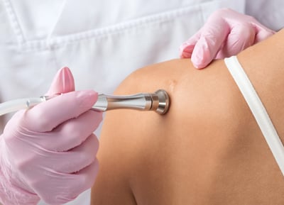
Mohs surgery is done on an outpatient basis in a procedure room with a nearby laboratory that allows the surgeon to examine the tissue after it is removed. The procedure, on average, requires less than four hours. However, it is not possible to predict how lengthy the procedure will be, as the extent of the skin cancer “roots” cannot be estimated in advance. The chart below walks you through each step of the Mohs surgery process:
Step 1 – To prepare you for surgery, the area to be treated is cleansed and injected with a local anesthetic. The anesthetic numbs the skin, so you won’t feel any discomfort during the procedure.
Step 2 – Once the anesthetic has taken effect, the Mohs surgeon uses a scalpel to remove the visible cancer, along with a thin layer of additional tissue. This procedure takes only a few minutes.
Step 3 – The tissue, once removed, is then divided into sections. The Mohs surgeon then color codes each of these sections with dyes and makes reference marks on the skin to show the source of these sections. A Mohs map of the site is then drawn.
Step 4 – The surgeon then brings this tissue to the laboratory for analysis. This portion of the procedure typically takes the longest amount of time, often requiring one hour or more to complete. The tissue is frozen on a cryostat, and the technician removes very thin slices from the entire edge and undersurface. These slices are then placed on slides and stained for examination under the microscope.
Step 5 – The Mohs surgeon carefully examines the entire undersurface and complete edge of the specimen. All microscopic “roots” of the cancer are precisely identified and pinpointed on the Mohs map. Upon microscopic examination, if residual cancer is found, the Mohs surgeon utilizes the Mohs map to direct the removal of additional tissue. Additional tissue is removed only where cancer is present.
Step 6 – This process is repeated as many times as necessary to locate and remove any remaining cancerous areas within the tissue specimen. Local anesthetic can be re-administered as necessary.
Step 7 – The removal process stops when there is no longer any evidence of cancer remaining in the surgical site. Because Mohs surgery removes only tissue containing cancer, it ensures that the healthy tissue is kept intact. Bleeding will be stopped with an electrocautery. You will be able to hear a buzzing noise and notice a distinct smell as it cauterizes the blood vessels. After all the cancer has been removed, you and your Mohs surgeon can decide on how to repair the wound. Most defects are repaired with reconstructive surgery which includes flaps, grafts, and linear closures. The reconstruction type will vary depending on the location, size, and depth of the wound.
While Mohs surgery can be a scary treatment to walk into, your Mohs surgeon will ensure that at each step in the process you are comfortable and understanding what is happening next. As a patient, never be afraid to ask a question of your Mohs surgeon – they are here to make sure your experience is a positive one.
Skin Struggles?
If you are struggling with acne or other skin issues and don’t know where to turn, the skin health experts at Forefront Dermatology are ready to help. To find the Forefront dermatologist nearest you, visit the locations page today.

Book an appointment with your trusted, local dermatologist.

How to Properly Apply Sunscreen

Bemotrizinol: What Does This New Sunscreen Ingredient Mean for the U.S.?
AI Skin Apps vs. Dermatologists: Why Professional Diagnosis Still Matters

Sun Safety Simplified: The Dermatologist-Approved Way
The Top 5 Aesthetic Treatments to Restore Your Look After Skin Cancer Treatment

Can Botox or Dermal Fillers Be Used After Skin Cancer Treatment?