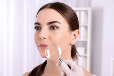
There is a moment, early in many dermatology visits, when a patient feels the air shift. The doctor has been studying a mole or a patch of skin with quiet concentration. Maybe they reach for a magnifying dermatoscope and look a little longer than expected. The patient is already running through the next steps in their head — biopsy, waiting, results, what if it’s something — before any of those words have been said out loud.
For decades, the path from “let’s take a closer look” to “this is what it is” has run almost exclusively through the skin biopsy. And while biopsies remain one of the most important diagnostic tools in dermatology, a newer technology called reflectance confocal microscopy (RCM) is opening up another pathway.
What is reflectance confocal microscopy (RCM)?
RCM is a non-invasive imaging technique that lets dermatologists gather diagnostic information before any tissue is removed, sometimes ruling out the need for a biopsy, and otherwise guiding a more precise one.
Most people are familiar with a regular microscope: the kind that examines a slide of tissue after it has been removed from the body. RCM is a microscope that can look into the skin while it is still on the patient and produce images at a level of detail close to what a pathologist sees on a slide. In fact, RCM imaging is so detailed that it is sometimes referred to as an optical biopsy.
What to expect during an RCM procedure
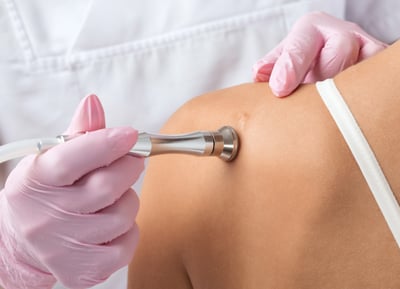
RCM works using a low-powered laser that is safe for both the patient’s skin and the operator’s eyes.
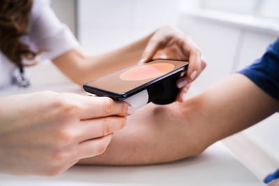
First, a small amount of oil is dabbed onto the skin to help the laser pass through clearly. A small metal ring with a glass window is placed over the spot, and the microscope’s lens is snapped onto the ring magnetically.
The laser shines through the lens and into the skin, and a sensor captures the light that bounces back from a single, precisely focused point. By collecting only that focused light and filtering out the rest, the device builds a sharp image of the skin one tiny layer at a time.The dermatologist sees live images of the skin’s individual cells on a nearby monitor.
The procedure is painless and non-invasive. A patient who walks in worried about a suspicious spot can sometimes walk out the same visit with real diagnostic information that does not require a biopsy.
RCM vs. Skin Biopsy
In many situations, a biopsy will still be the right next step, and will remain the diagnostic gold standard for the foreseeable future. However, RCM helps dermatologists determine whether a biopsy is needed, when one is needed, and where to take it.
The research on RCM’s usefulness is increasingly compelling. A large randomized clinical trial published in JAMA Dermatology found that using RCM alongside visual dermatological exams reduced unnecessary biopsies by more than 43%, while still identifying invasive melanomas accurately. Even patients who were ultimately diagnosed with skin cancer benefited from more precise biopsy targeting using information gathered during RCM.
Translated out of the clinical language, that means something simple and meaningful: fewer scars, fewer anxious weeks waiting for results, and fewer biopsies done “just to be sure” on spots that turn out to be nothing.
Benefits of RCM
RCM is particularly valuable in situations where a traditional biopsy carries higher emotional or cosmetic stakes:
- Lesions on the face, where a biopsy can leave a visible scar and where many of the trickiest pigmented lesions tend to appear.
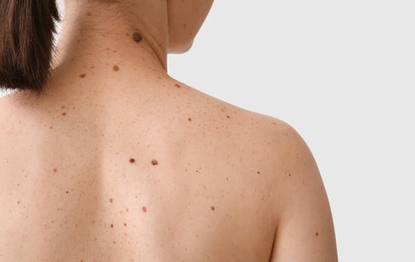
- Patients with many moles, whose dermatologists often have to choose between biopsying every borderline spot or watching them closely over time.
- Spots that are tricky to interpret with the naked eye or a dermatoscope, including lesions without the typical color cues and ones that look like they’re partially healing or changing on their own.
- Mapping the edges of certain skin cancers before surgery, so the doctor knows where the affected tissue truly ends.
What RCM can help diagnose
Research and real-world use have shown RCM to be especially helpful in evaluating:
- Melanoma, including lentigo maligna, a slow-growing form of melanoma that often appears on sun-damaged skin of the face, where it can be especially difficult to distinguish from harmless age spots.
- Basal cell carcinoma, the most common form of skin cancer, where RCM can often confirm the diagnosis and help map the borders before treatment.
- Squamous cell carcinoma, another common skin cancer that RCM can help identify in its earlier stages.
- Unusual moles and pigmented lesions, particularly the borderline cases where it isn’t immediately clear whether a spot is benign or concerning.
- Inflammatory skin conditions such as psoriasis and eczema, where RCM can sometimes clarify what’s happening at the cellular level without a biopsy.
- Treatment monitoring, including checking how well certain non-surgical skin cancer treatments are working over time.
What RCM cannot do
Though the RCM delivers numerous benefits, the technology does have limitations.
RCM can only see into the upper layers of the skin: roughly down to the papillary dermis. Anything deeper than that requires a traditional biopsy. And in certain locations, such as the inside of the ear, the inner corner of the eye, or anywhere the device cannot sit flat, the technology simply does not work as well.
The images also take real expertise to interpret; accurate interpretation is a learned skill that not every dermatologist has experience with. For patients, that means it’s worth seeking out a practice with dermatologists specifically trained in reading RCM images.
RCM is not a replacement for the careful, layered approach that dermatology has always relied on. It is an addition to it.
Learn more about RCM
Staying at the leading edge of dermatology isn’t about chasing every new device. It’s about choosing the ones that genuinely make care better: fewer unnecessary procedures, more confident diagnoses, and less time spent waiting.
Reflectance confocal microscopy is one of those tools. It will not be the right answer for every lesion or every patient, and it does not replace the foundational role of biopsy in diagnosing skin cancer and many other skin conditions. But for the right person at the right moment, it can mean walking out of the office with information instead of another appointment, and clarity instead of uncertainty.
Concerned about a spot on your skin? Find a Forefront location near you to get a clear answer about what’s next.

Book an appointment with your trusted, local dermatologist.
What Does Skin Cancer Look Like? A Dermatologist’s Guide to Early Signs, Warning Symptoms, and When to Get Checked

How to Properly Apply Sunscreen

Bemotrizinol: What Does This New Sunscreen Ingredient Mean for the U.S.?
AI Skin Apps vs. Dermatologists: Why Professional Diagnosis Still Matters

Sun Safety Simplified: The Dermatologist-Approved Way
The Top 5 Aesthetic Treatments to Restore Your Look After Skin Cancer Treatment